Unidades Centrales de Apoyo a la Investigación Biomédica
Microscopía óptica avanzada


La Unidad de Microscopía Óptica Avanzada de IMIBIC pone a disposición de los investigadores de IMIBIC y de Instituciones públicas o privadas, equipamiento especializado con una amplia variedad de microscopios que cubren un amplio espectro de aplicaciones y técnicas empleadas en investigación biomédica.
El objetivo de la Unidad es dar un soporte integral a los investigadores proporcionando asesoramiento y soporte científico-técnico sobre diseño experimental, preparación de muestras, procesamiento y análisis de imágenes, e interpretación de resultados.
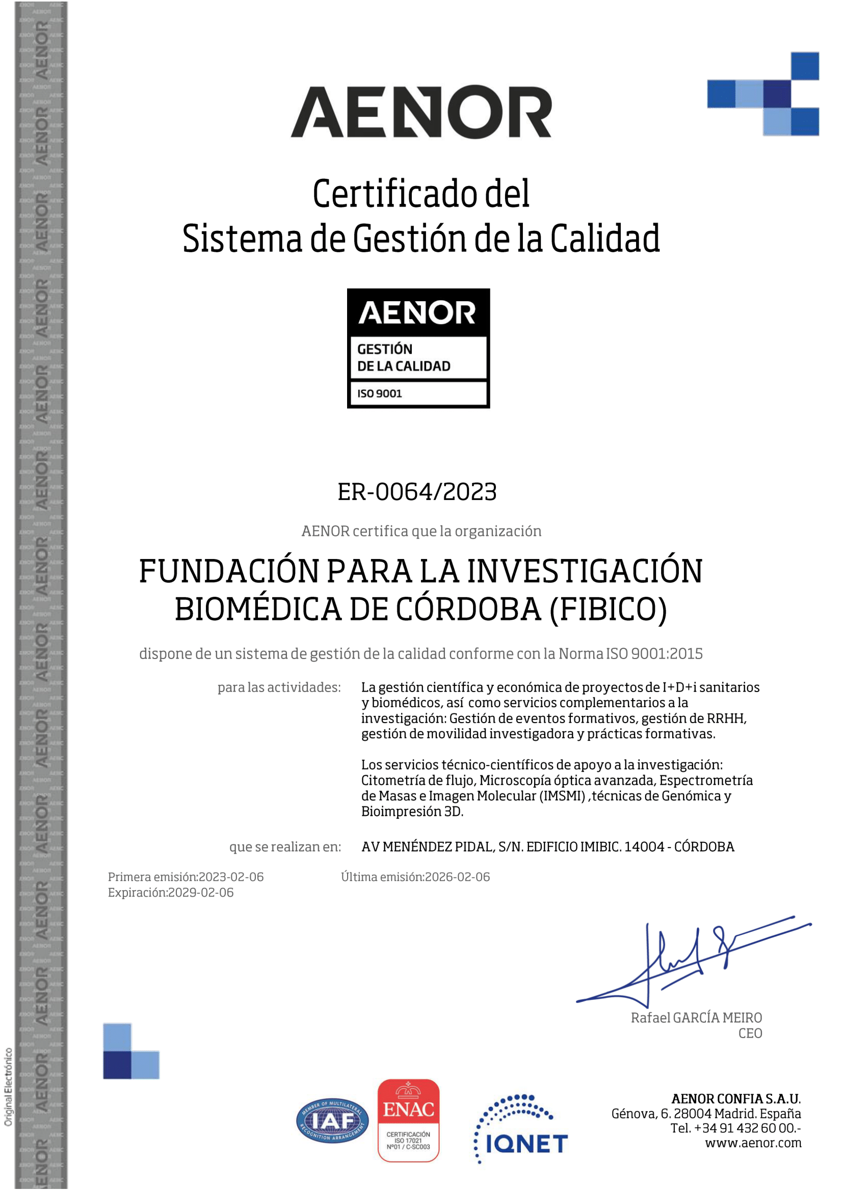
La unidad de Microscopía óptica avanzada dispone de un Sistema de Gestión de Calidad conforme a la Norma de Calidad ISO 9001:2015.
Personal

Gema García Jurado, PhD
Responsable de Unidad. Especialista Técnico de Microscopía
- gema.garcia@imibic.org
- microscopia@imibic.org
- (+34) 957 213 753 (Corporativo 3753)
- 609 15 77 90

Alvaro Carrasco Carmona
Especialista Técnico Senior
Equipamiento e infraestructuras
Microscopía Confocal
Microscopía de Fluorescencia y campo claro
Microdisección láser
Estación y software para análisis de imagen
Microscopio CONFOCAL ESPECTRAL LSM 710 (Carl Zeiss)
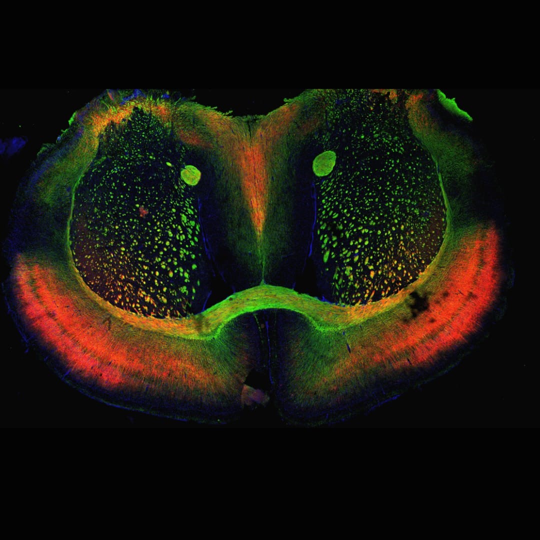
La Microscopía confocal espectral tiene la capacidad de controlar la profundidad de campo eliminando o reduciendo las señales luminosas procedentes de fuera del plano focal, incrementando así la resolución y contraste de las imágenes para obtener secciones ópticas seriadas de la muestra, y su posterior reconstrucción 3D.
-
Microscopio Axio Observer.Z1 invertido motorizado con sistema de iluminación para campo claro, DIC y epifluorescencia.
-
Láseres de excitación: láser Ar (458, 488, 514 nm), láser HeNe (543 nm), láser HeNe (594 nm), láser HeNe (633 nm) y láser diodo (405 nm).
-
Detección: 3 canales de detección espectral de fluorescencia con PMTs, y 1 canal para luz transmitida con PMT y capacidad DIC.
-
Sistema de incubación celular integrado XL.
-
Configuración de FILTROS DE FLUORESCENCIA:

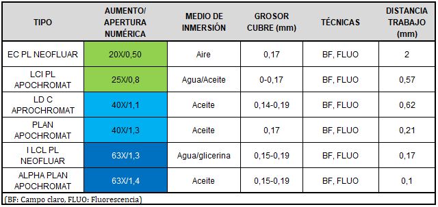
OBJETIVOS:
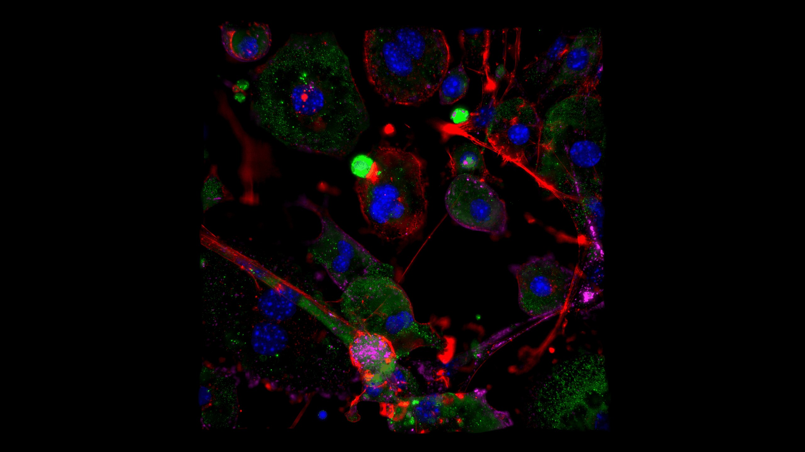
Microscopio de fluorescencia LEICA THUNDER IMAGER 3D ASSAY
-
Microscopio invertido DMi8 completamente motorizado.
-
Software de imagen Las X con aclaramiento computacional THUNDER El Módulo de software “Thunder” permite la obtención de imágenes libres de la señal proveniente de fuera del plano de foco, mediante deconvolución a tiempo real.
-
ILUMINACIÓN DE FLUORESCENCIA con 5 LEDs que generan un espectro continuo entre 390 y 680 nm.
-
CÁMARA LEICA monocromática K5 de alta sensibilidad. Sensor sCMOS B/N de 2048x2048 píxeles, tamaño del píxel 6,5 micras.
-
Configuración de FILTROS DE FLUORESCENCIA:

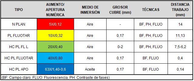
OBJETIVOS:
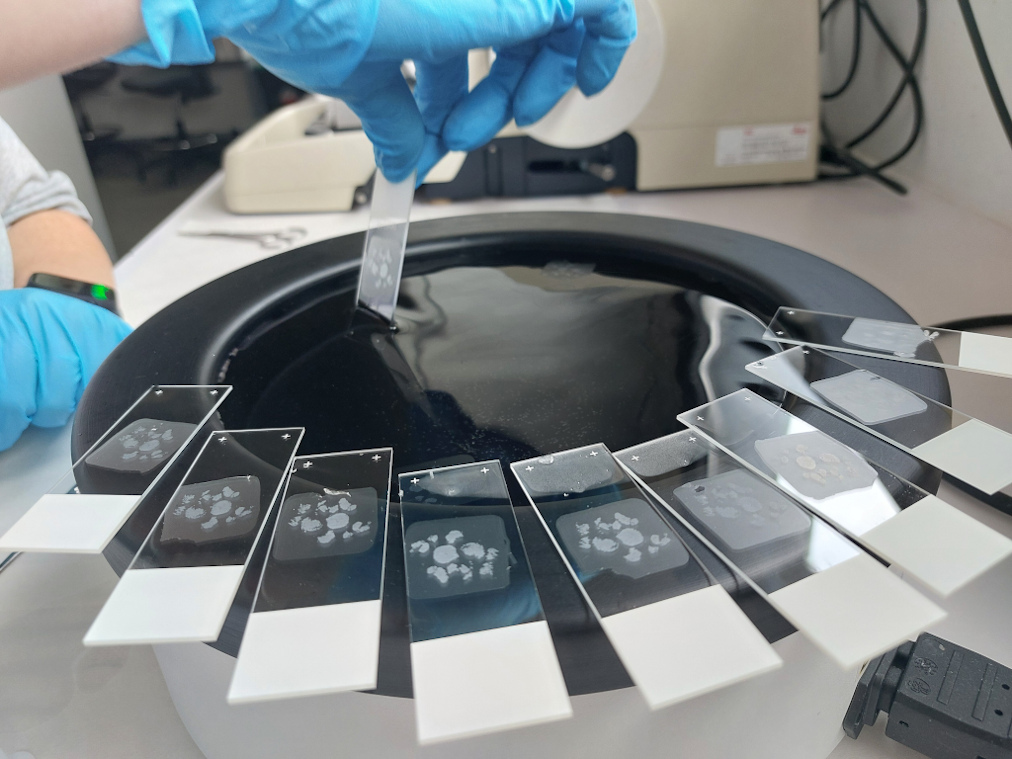
MICRODISECTOR Láser ZEISS PALM Microbeam
Sistema de microdisección láser PALM de ZEISS para la selección dirigida de muestras en preparaciones celulares y tisulares. El sistema de microdisección utiliza un láser de estado sólido de 355nm focalizado a través del objetivo para cortar, aislar y catapultar la muestra seleccionada sin contacto con una precisión inferior a 1μm.
-
Tipos de muestras para diseccionar: muestras histológicas en fresco y fijadas, cultivos celulares (célula viva), Cytospin, biofilms, material forense, etc. Para estudios in vivo consta de un sistema de incubación sobre platina para mantener atmósfera controlada, pH y concentración de CO2.
-
El láser permite trabajar tanto con luz transmitida en campo claro, con contraste de fases y también realizar microdisección en fluorescencia con muestras marcadas con cromóforos fluorescentes.
-
Consta de 2 cámaras digitales: Cámara color Axiocam 305 y cámara monocroma Axiocam 305.
-
Software de control del microdisector: PALM RoboSoftware.
-
Softwares de adquisición y análisis de imágenes Axiovision 4.9.1 y Zen Blue 3.4.
-
Configuración de FILTROS DE FLUORESCENCIA:

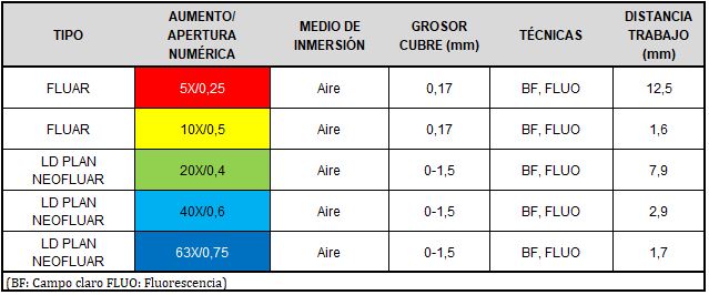
OBJETIVOS:
Microscopio de FLUORESCENCIA Axio Vert.A1 (Carl Zeiss).
-
Microscopio invertido con sistema de iluminación para campo claro y epifluorescencia.
-
Cámara CCD monocroma AxioCam ICm1-1.4 Mpx.
-
Software de adquisición y análisis ZEN 2 (Zeiss).
-
Configuración de FILTROS DE FLUORESCENCIA:

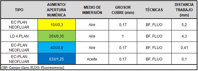
OBJETIVOS:
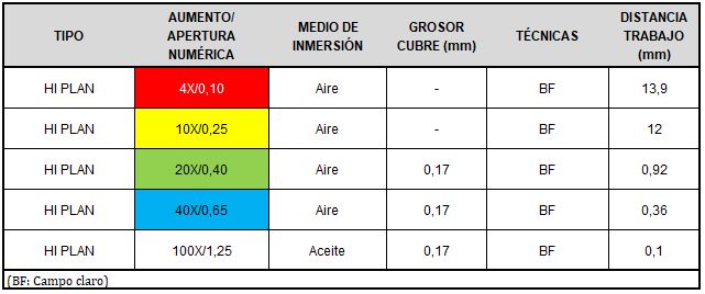
Microscopio Óptico Leica DM2000 LED
-
Microscopio vertical con iluminación LED para campo claro.
-
Cámara digital HD color.
OBJETIVOS:
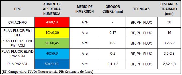
Microscopio Óptico NikonEclipse Ti-S
-
Microscopio invertido con sistema para campo claro y contraste de fases.
OBJETIVOS:
Estación de trabajo 3D
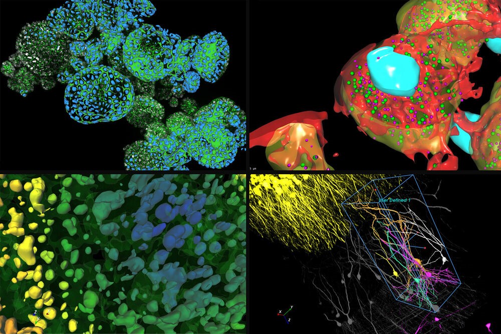
La estación de trabajo es una potente estación para el análisis y procesado de imágenes 3D. Esta estación dispone del software de análisis AIVIA CellBio. Es un software avanzado de visualización y análisis de imagen basado en inteligencia artificial. Este software está especialmente orientado al análisis celular, permitiendo el estudio de múltiples compartimentos relacionados entre sí, tanto en 2D como en 3D.
Otros programas disponibles: FIJI, LAS X, Zen Blue, QuPath.
Servicios
- Microscopía laser confocal.
- Microscopía automatizada para campo claro y fluorescencia.
- High content screening.
- Microscopía “in vivo” 3D.
- Procesado instantáneo de imágenes (computational clearing).
- Microdisección láser.
- Imágenes espectrales / Linear Unmixing.
- Procesado y análisis de imágenes.
Servicios