Unidades Centrales de Apoyo a la Investigación Biomédica
Imagen Preclínica

El servicio de imagen preclínica (SIP) es una instalación de vanguardia que ofrece imágenes in vivo y no
invasivas de la estructura y función de los órganos y tejidos del cuerpo de modelos animales
preclínicos.
Este equipamiento se encuentra dentro del animalario del IMIBIC, el cual cuenta con unas modernas
instalaciones de aproximadamente 800 m² y capacidad para estabulación y manejo experimental de
>3500 roedores, hallándose en una localización ideal para proporcionar trabajo interdisciplinario en
biología cardiovascular, oncología, neurociencia y fisiología, entre otras áreas.
Personal
Equipamiento e instalaciones
- PET/RM 3T, que constituye el único equipo combinado de resonancia magnética y PET disponible a nivel nacional, integra un equipo PET basado en la tecnología de cristal continuo LYSO de alta sensibilidad con 2 anillos de 8 detectores que permiten obtener campos de visión de 80 mm (transaxial) X 148 mm (axial), pudiendo llegar hasta 285 mm (axial) moviendo la cama, fotomultiplicadores de silicio compatibles con la RM, sensibilidad del 5 % pudiendo llegar al 9 % (norma NEMA) y resolución sub-milimétrica (≤ 0,7 mm); el equipo de RM es de 3 Tesla y boca de imán de 18 cm con tecnología cryogen free, sin necesidad de línea de quench, con una homogenidad del imán de ± 0,1 ppm (DSV 5 cm) y ± 0,05 ppm (DSV 3,5 cm), necesaria para trabajar con secuencias de difusión y en espectroscopía localizada y un con un sistema de gradientes capaz de proporcionar 450 mT/m. El PET/RM presenta un sistema automático de posicionamiento del animal tanto para la modalidad PET como en RM y pantalla touch screen con cámara integrada para facilitar las labores de posicionamiento y seguimiento.
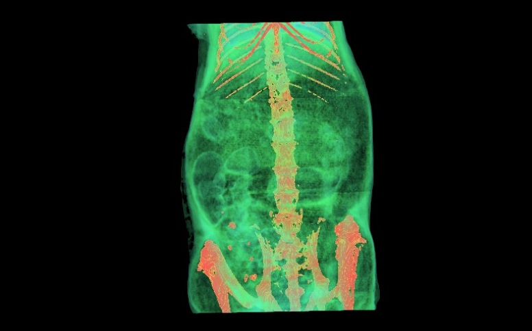
- μCT (modelo SkyScan 1176), que constituye un sistema de tomografía de rayos X (fuente de 90 kV) con un resolución espacial 3D de menos de 10 μm y un sistema de gating respiratorio para sincronización prospectiva o retrospectiva de movimientos respiratorios durante la adquisición de la imagen. El sistema cuenta con una cámara refrigerada de rayos X de 12 bits, de 11 Mp con distorsión corregida (4000x2670) con acoplamiento de fibra óptica para centelleador. Capacidad de escaneo de cuerpo entero de rata y ratón: diámetro de escaneo 68 mm o 35 mm y longitudinal de hasta 200 mm por múltiples escáneres conectados de forma automática. Este equipo μCT es capaz de llevar a cabo ensayos longitudinales con una exposición de radiación inferior a 10 mGy además de co-registro de imágenes con el sistema PET o la RM.
Servicios
- Asesoramiento en el diseño experimental, elección de los equipos y supervisión de los procedimientos.
- Asesoramiento durante la adquisición y el procesamiento de imágenes mediante resonancia magnética (RM), micro-tomografía computarizada (micro-TAC) y tomografía por emisión de positrones (PET).
- Aplicaciones específicas:
- Análisis estructural de tejidos blandos y de composición corporal (compartimentación).
- Análisis de estructuras mediante difusión (diffusion weighted image[DWI]).
- Análisis de actividad cerebral mediante fMRI.
- Análisis estructurales.
- Análisis de la actividad metabólica (tejido sano y tumoral).

